Details of Host Protein
| Host Protein General Information (ID: PT0340) | |||||||||
|---|---|---|---|---|---|---|---|---|---|
| Protein Name |
DNA damage-binding protein 1 (DDB1)
|
Gene Name |
DDB1
|
||||||
| Host Species |
Homo sapiens
|
Uniprot Entry Name |
DDB1_HUMAN
|
||||||
| Protein Families |
DDB1 family
|
||||||||
| Subcellular Location |
Cytoplasm Nucleus
|
||||||||
| External Link | |||||||||
| NCBI Gene ID | |||||||||
| Uniprot ID | |||||||||
| Ensembl ID | |||||||||
| HGNC ID | |||||||||
| Function in Host |
Protein, which is both involved in DNA repair and proteinubiquitination, as part of the UV-DDB complex and DCX (DDB1-CUL4-X-box) complexes, respectively. Core component of the UV-DDB complex (UV-damaged DNA-binding protein complex), a complex that recognizes UV-induced DNA damage and recruit proteins of the nucleotide excisionrepair pathway (the NER pathway) to initiate DNA repair. The UV-DDB complex preferentially binds to cyclobutane pyrimidinedimers (CPD), 6-4 photoproducts (6-4 PP), apurinic sites and shortmismatches. Also functions as a component of numerous distinctDCX (DDB1-CUL4-X-box) E3 ubiquitin-protein ligase complexes whichmediate the ubiquitination and subsequent proteasomal degradation oftarget proteins. The functional specificity of the DCX E3 ubiquitin-protein ligasecomplex is determined by the variable substrate recognition componentrecruited by DDB1. DCX (DDB2) (alsoknown as DDB1-CUL4-ROC1, CUL4-DDB-ROC1 and CUL4-DDB-RBX1) mayubiquitinate histone H2A, histone H3 and histone H4 at sites of UV-induced DNA damage. The ubiquitination of histones may facilitate theirremoval from the nucleosome and promote subsequent DNA repair. DCX (DDB2) also ubiquitinates XPC, which may enhance DNA-binding by XPCand promote NER. DCX (DTL) plays a role in PCNA-dependent polyubiquitination of CDT1 and MDM2-dependent ubiquitinationof TP53 in response to radiation-induced DNA damage and during DNAreplication. DCX (ERCC8) (the CSA complex) plays arole in transcription-coupled repair (TCR). The DDB1-CUL4A-DTL E3 ligase complex regulates the circadian clock function bymediating the ubiquitination and degradation of CRY1. DDB1-mediated CRY1 degradation promotes FOXO1 protein stability andFOXO1-mediated gluconeogenesis in the liver.
[1-15]
Click to Show/Hide
|
||||||||
| Related KEGG Pathway | |||||||||
| Hepatitis B | hsa05161 |
Pathway Map 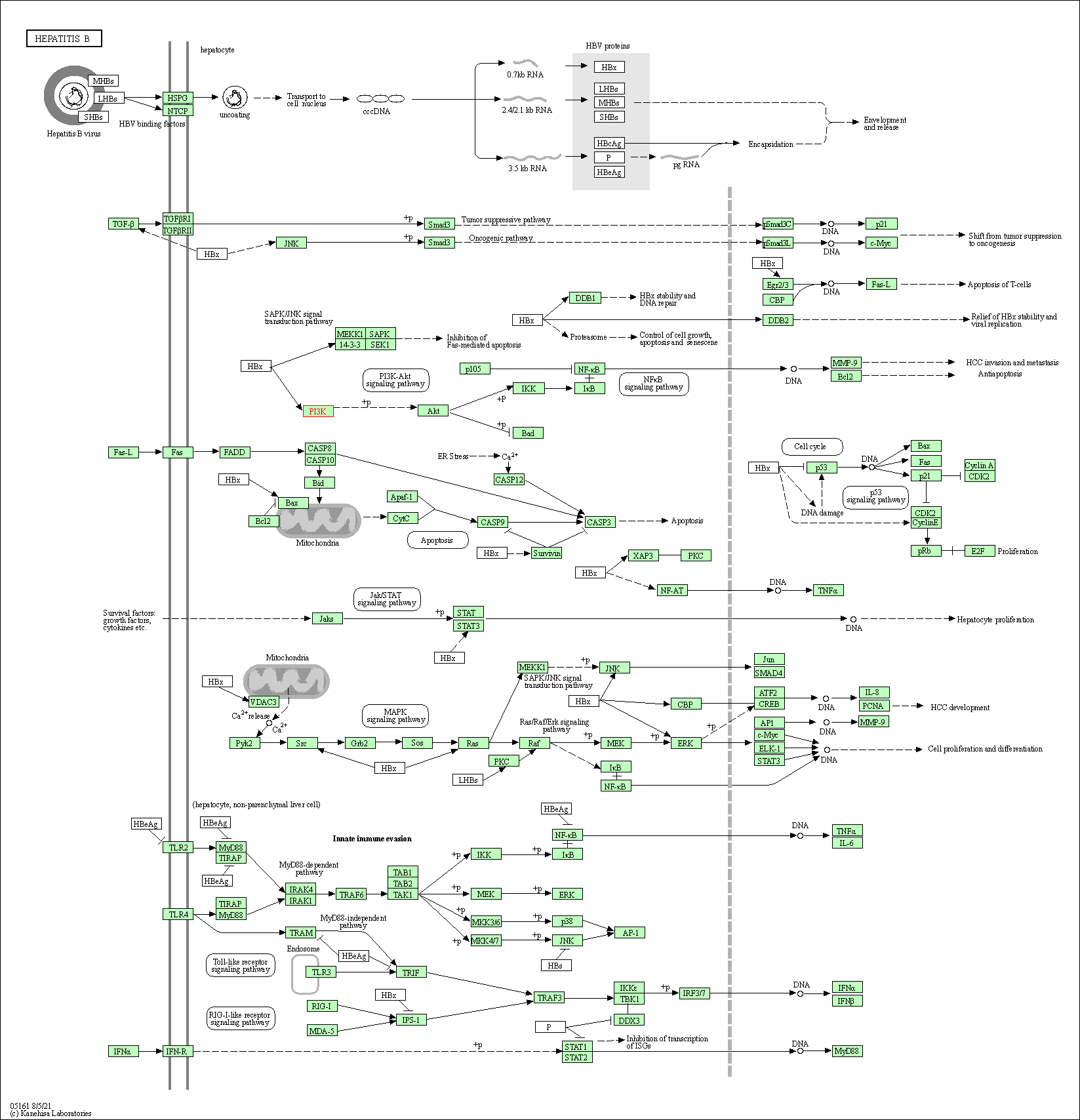
|
|||||||
| Human immunodeficiency virus 1 infection | hsa05170 |
Pathway Map 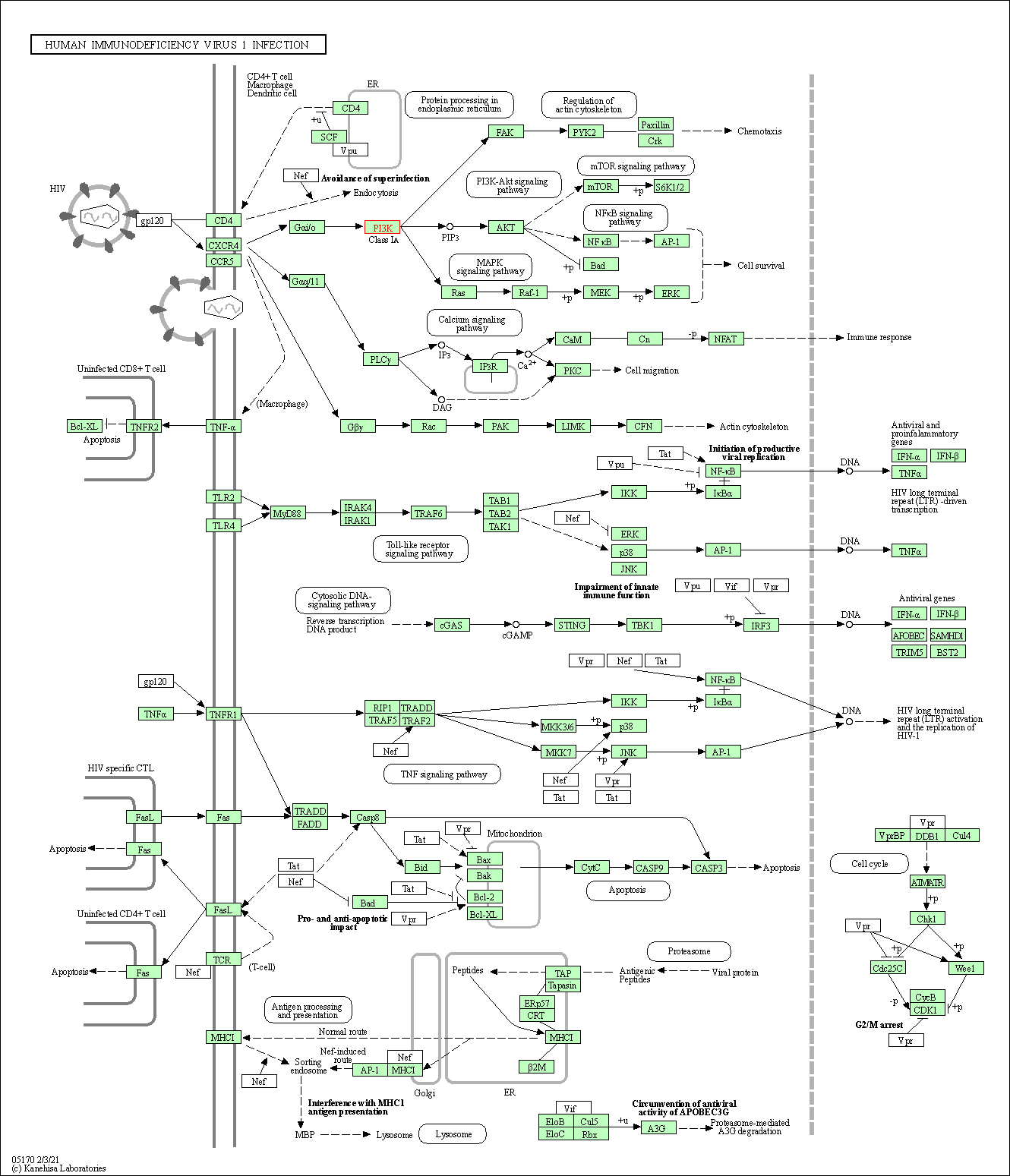
|
|||||||
| Viral carcinogenesis | hsa05203 |
Pathway Map 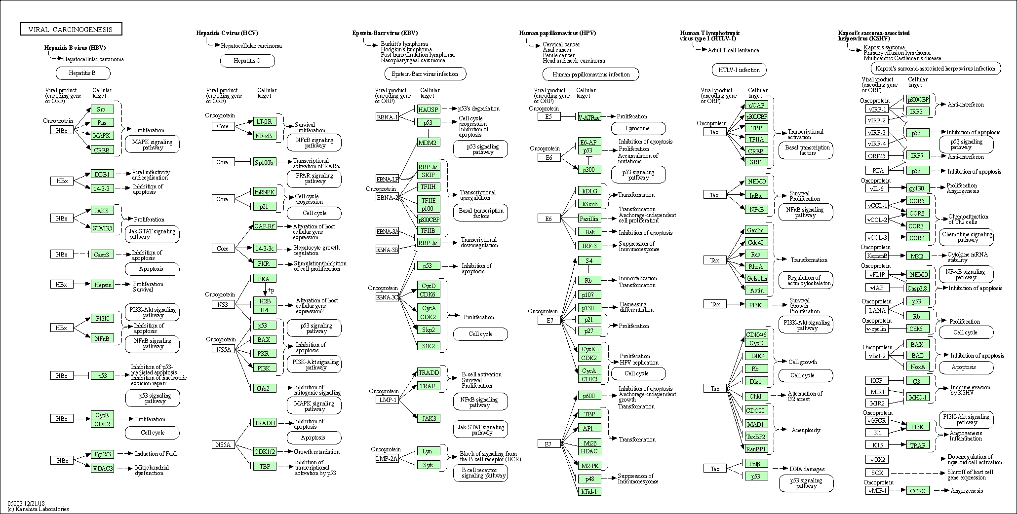
|
|||||||
| Nucleotide excision repair | hsa03420 |
Pathway Map 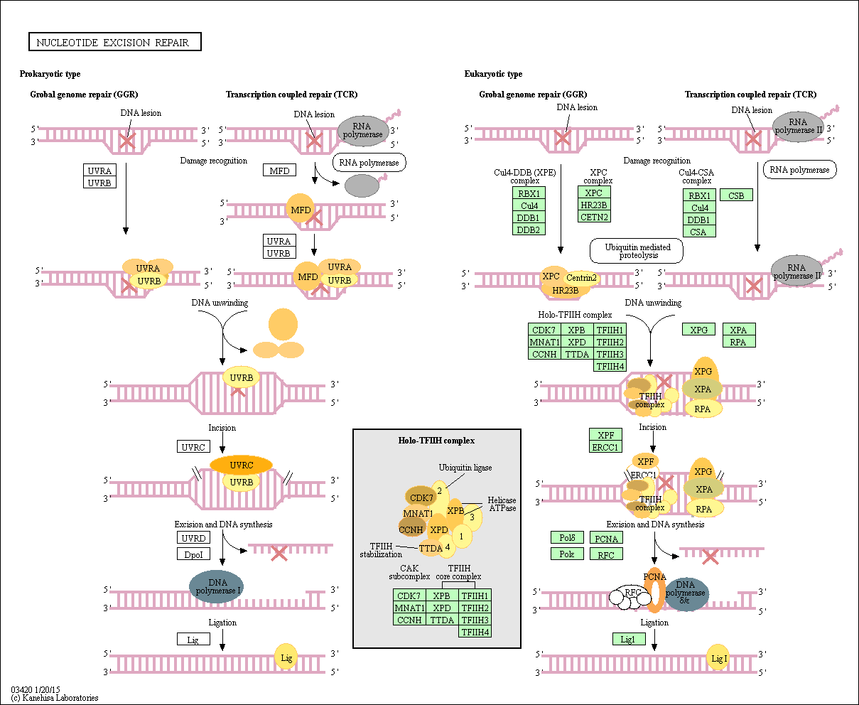
|
|||||||
| Ubiquitin mediated proteolysis | hsa04120 |
Pathway Map 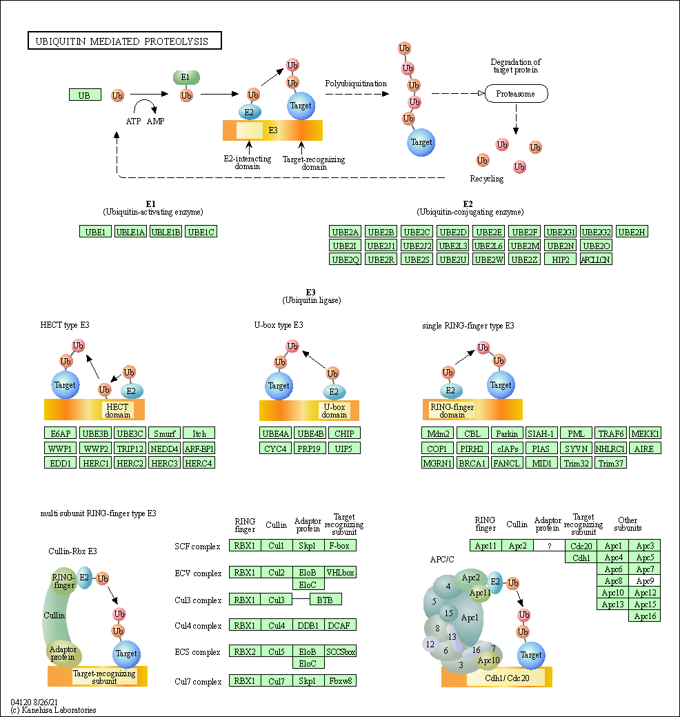
|
|||||||
| 3D Structure |
|
||||||||
| Full List of Virus RNA Interacting with This Protien | |||||||||
|---|---|---|---|---|---|---|---|---|---|
| RNA Region: ORF10 (hCoV-19/Not Specified Virus Strain ) | |||||||||
| RNA Region Details |
RNA Info
 Click to show the detail information of this RNA binding region
|
[16] | |||||||
| Strains Name |
hCoV-19/Not Specified Virus Strain
|
||||||||
| RNA Binding Region |
ORF10
|
||||||||
| Virus Name |
Severe acute respiratory syndrome coronavirus 2 (SARS-CoV-2)
|
||||||||
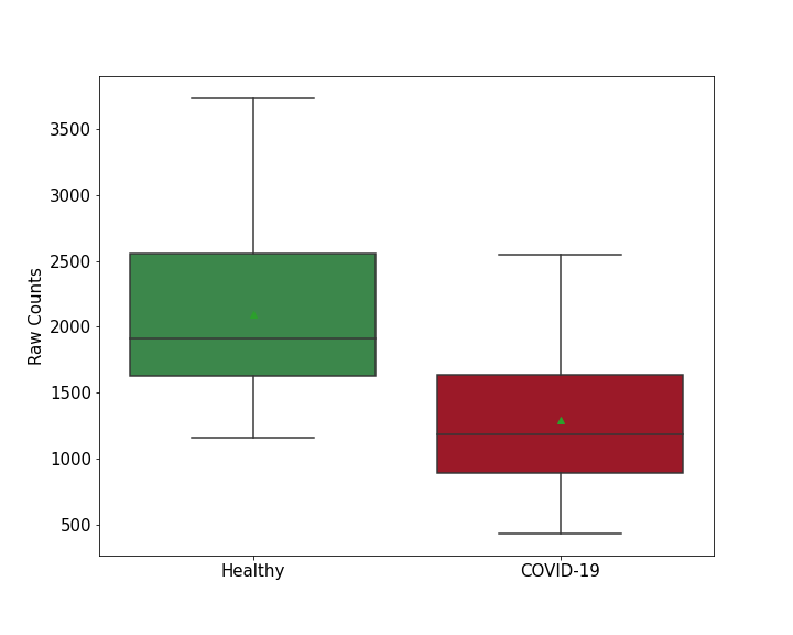
| Infection Cells | Calu-3 cells (Human Lung Cancer Cell) (CVCL_0609 ) | ||||||||
| Cell Originated Tissue | Liver | ||||||||
| Interaction Score | P-value < 0.05 | ||||||||
| Method Description | RNA pull-down assays; liquid chromatography with tandem mass spectrometry (LC-MS/MS); Wilcoxon test; MS2 affinity purification coupled with liquid chromatography-mass spectrometry (MAMS) | ||||||||