Details of Host Protein
| Host Protein General Information (ID: PT0584) | |||||||||
|---|---|---|---|---|---|---|---|---|---|
| Protein Name |
Cysteines of Keap1 (KEAP1 Cysteines)
|
Gene Name |
KEAP1
|
||||||
| Host Species |
Homo sapiens
|
Uniprot Entry Name |
KEAP1_HUMAN
|
||||||
| Protein Families |
KEAP1 family
|
||||||||
| Subcellular Location |
Cytoplasm Nucleus
|
||||||||
| External Link | |||||||||
| NCBI Gene ID | |||||||||
| Uniprot ID | |||||||||
| Ensembl ID | |||||||||
| HGNC ID | |||||||||
| Function in Host |
Substrate-specific adapter of a BCR (BTB-CUL3-RBX1) E3ubiquitin ligase complex that regulates the response to oxidativestress by targeting NFE2L2/NRF2 for ubiquitination. KEAP1 acts as a key sensor of oxidative and electrophilic stress: innormal conditions, the BCR (KEAP1) complex mediates ubiquitination anddegradation of NFE2L2/NRF2, a transcription factor regulatingexpression of many cytoprotective genes. In response to oxidative stress, differentelectrophile metabolites trigger non-enzymatic covalent modificationsof highly reactive cysteine residues in KEAP1, leading to inactivatethe ubiquitin ligase activity of the BCR (KEAP1) complex, promotingNFE2L2/NRF2 nuclear accumulation and expression of phase II detoxifyingenzymes. In response to selective autophagy, KEAP1 is sequestered in inclusion bodies following its interaction withSQSTM1/p62, leading to inactivation of the BCR (KEAP1) complex andactivation of NFE2L2/NRF2. The BCR (KEAP1) complexalso mediates ubiquitination of SQSTM1/p62, increasing SQSTM1/p62sequestering activity and degradation. The BCR (KEAP1) complex also targets BPTF and PGAM5 for ubiquitination and degradationby the proteasome.
[1-7]
Click to Show/Hide
|
||||||||
| Related KEGG Pathway | |||||||||
| Ubiquitin mediated proteolysis | hsa04120 |
Pathway Map 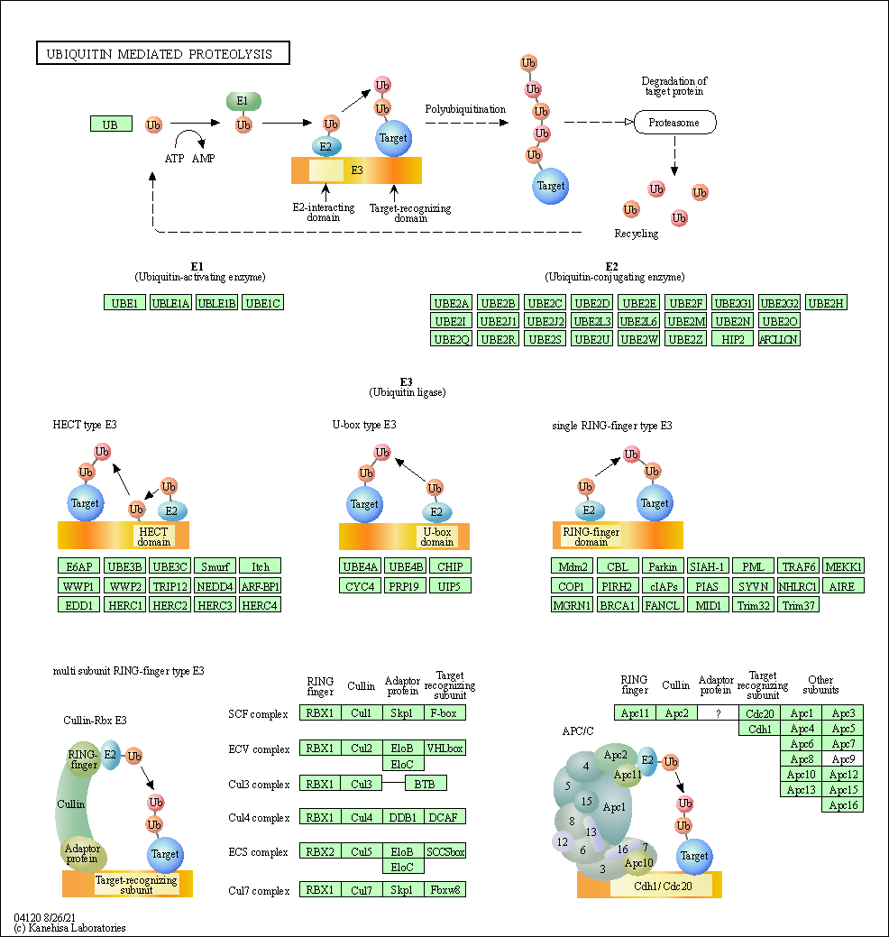
|
|||||||
| Pathways in cancer | hsa05200 |
Pathway Map 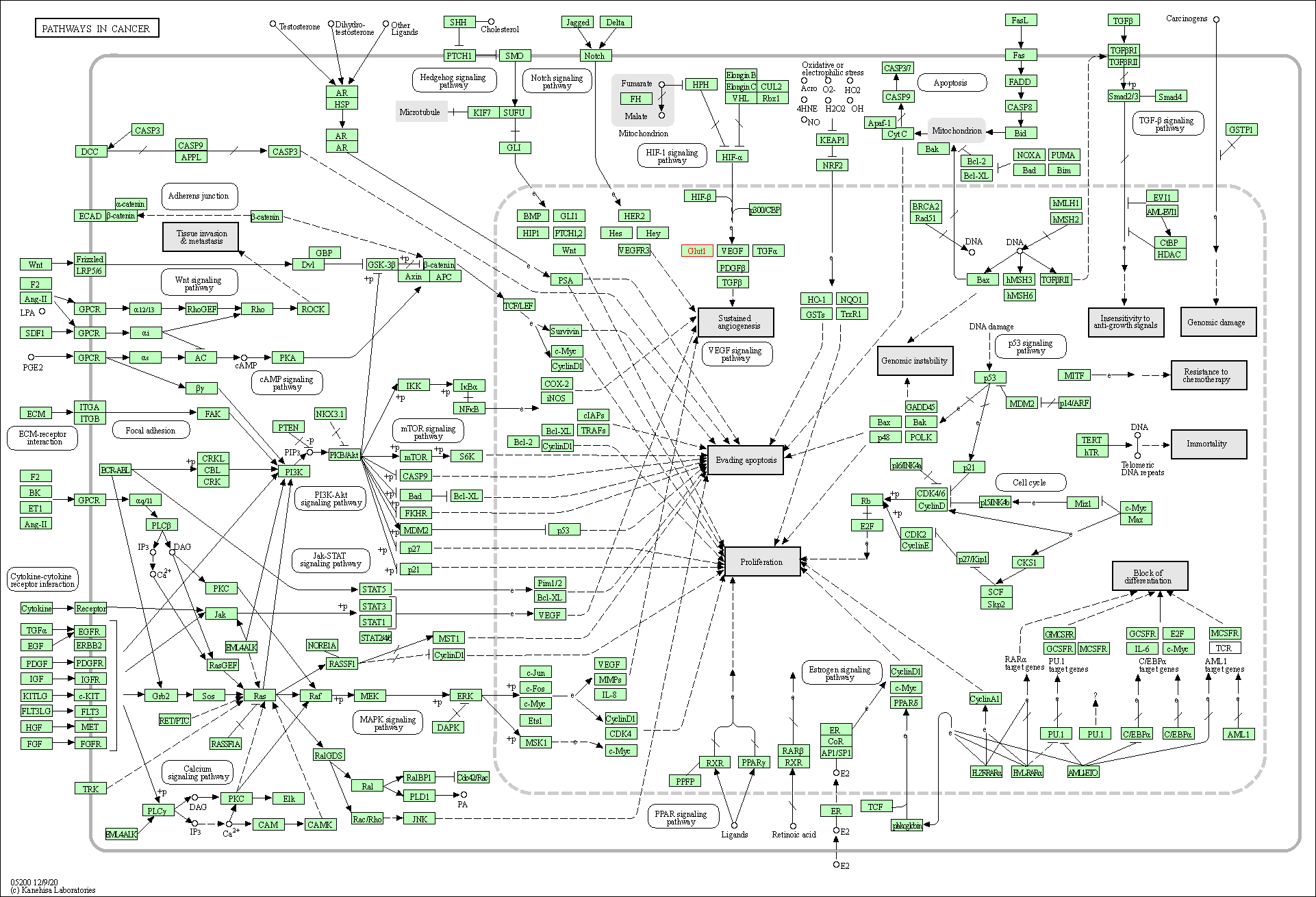
|
|||||||
| Chemical carcinogenesis - reactive oxygen species | hsa05208 |
Pathway Map 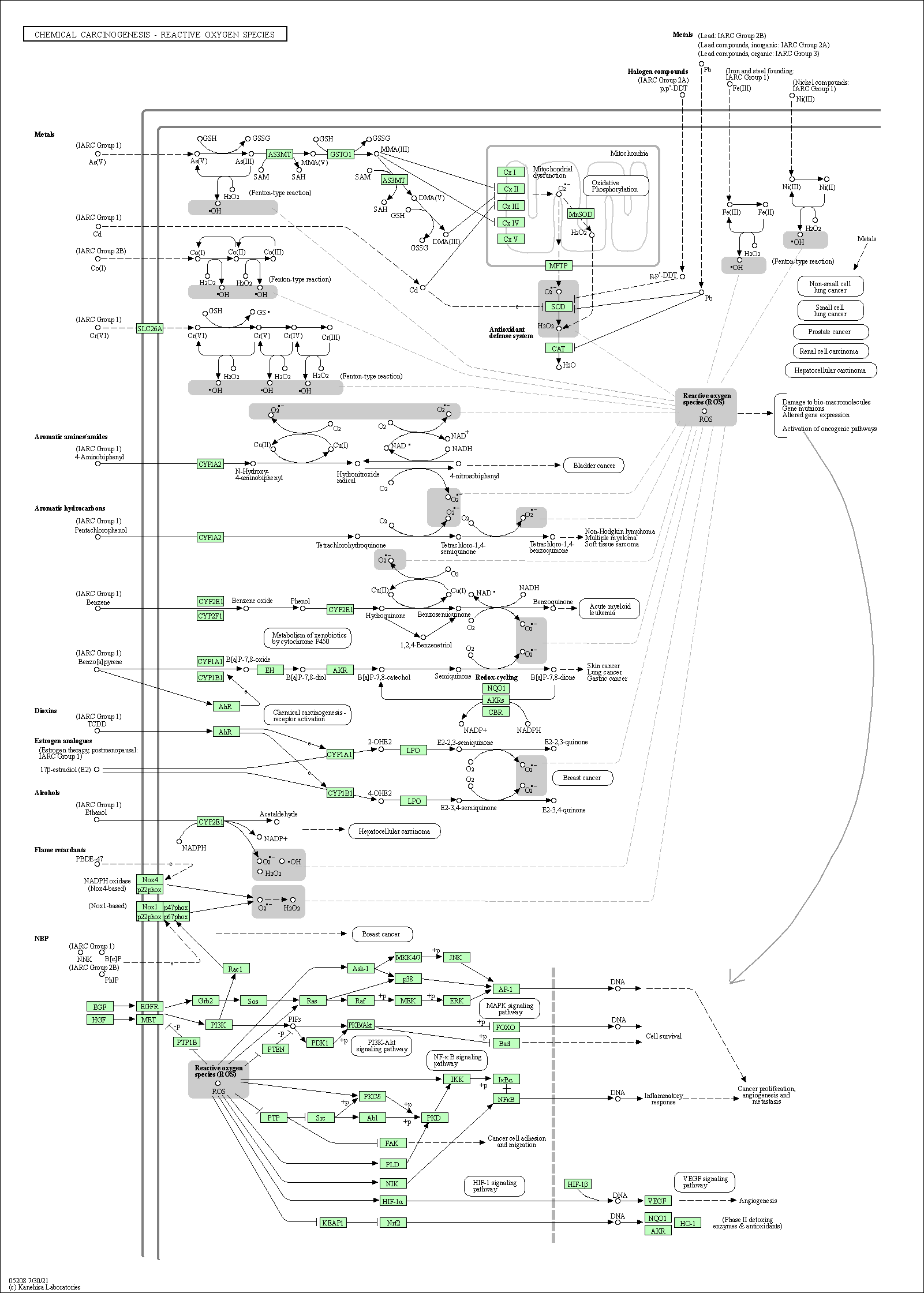
|
|||||||
| Hepatocellular carcinoma | hsa05225 |
Pathway Map 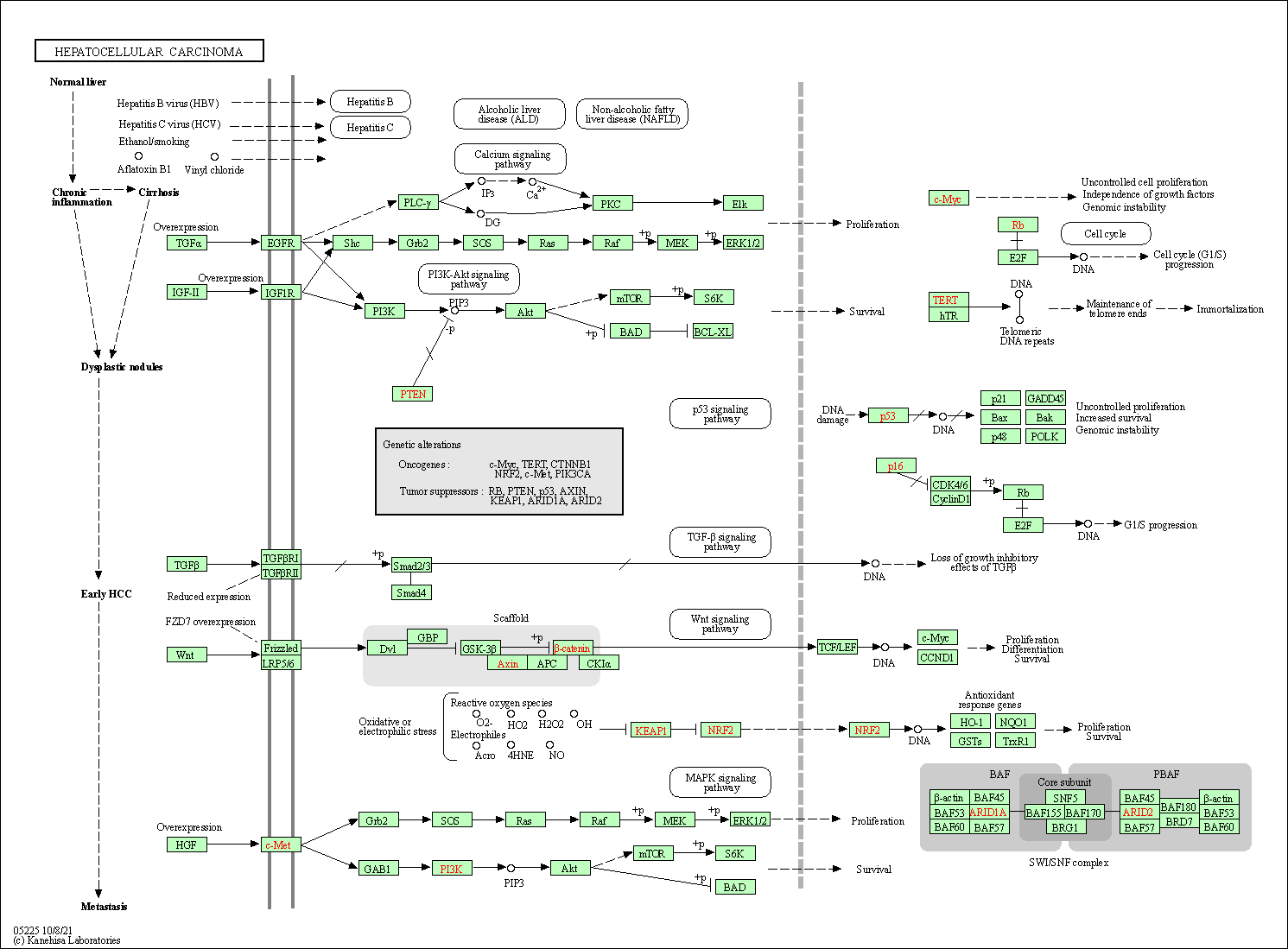
|
|||||||
| Fluid shear stress and atherosclerosis | hsa05418 |
Pathway Map 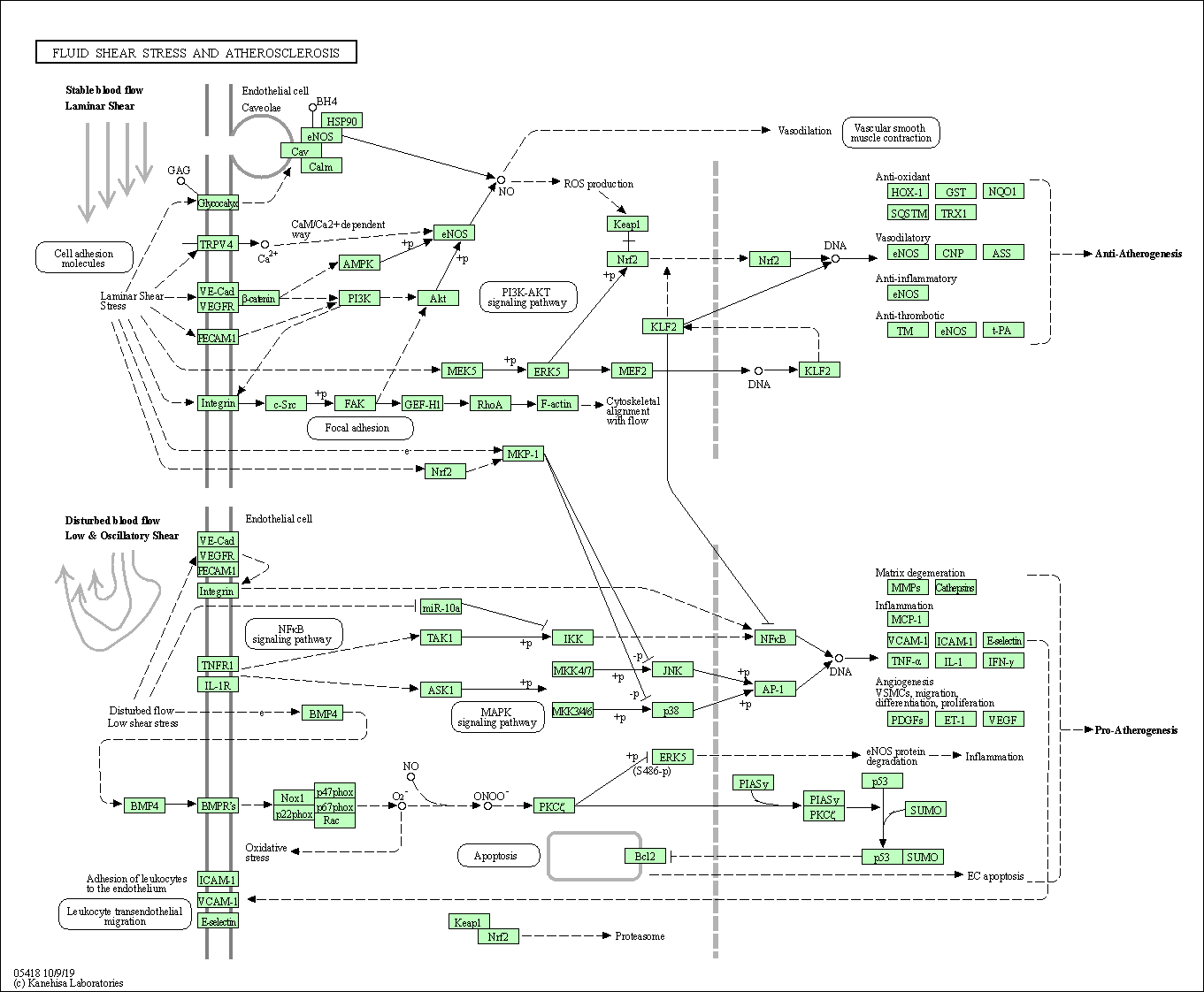
|
|||||||
| Parkinson disease | hsa05012 |
Pathway Map 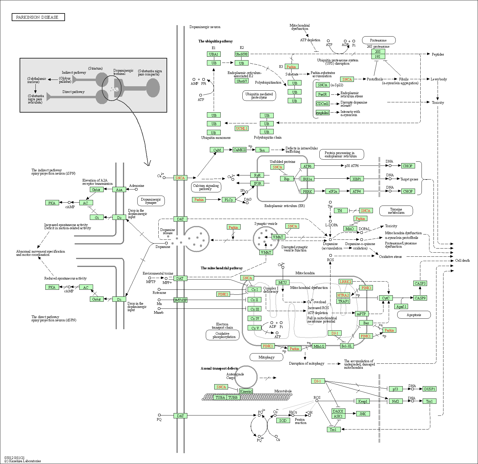
|
|||||||
| 3D Structure |
|
||||||||
| Function of This Protein During Virus Infection | |||||||||
|---|---|---|---|---|---|---|---|---|---|
| Virus Name | SARS-COV-2 | Protein Function | Anti-viral | [8] | |||||
| Infected Tissue | Lung | Infection Time | 7-9 Days | ||||||
| Infected Cell | Calu-3 Cells (Human epithelial cell line) | Cellosaurus ID | CVCL_0609 | ||||||
| Method Description | To detect the role of host protein KEAP1 in viral infection, KEAP1 protein knockout Calu-3 Cells were infected with SARS-COV-2 for 7 - 9 Days , and the effects on infection was detected through CRISPR-based genome-wide gene-knockout screen. | ||||||||
| Results | It is reported that knockout of KEAP1 increases SARS-CoV-2 RNA levels compared with control group. | ||||||||
| Full List of Virus RNA Interacting with This Protien | |||||||||
|---|---|---|---|---|---|---|---|---|---|
| RNA Region: ORF10 (hCoV-19/Not Specified Virus Strain ) | |||||||||
| RNA Region Details |
RNA Info
 Click to show the detail information of this RNA binding region
|
[9] | |||||||
| Strains Name |
hCoV-19/Not Specified Virus Strain
|
||||||||
| RNA Binding Region |
ORF10
|
||||||||
| Virus Name |
Severe acute respiratory syndrome coronavirus 2 (SARS-CoV-2)
|
||||||||
| Infection Cells | Huh7 cells (human liver cell line) (CVCL_0336 ) | ||||||||
| Cell Originated Tissue | Liver | ||||||||
| Interaction Score | P-value < 0.05 | ||||||||
| Method Description | RNA pull-down assays; liquid chromatography with tandem mass spectrometry (LC-MS/MS); Wilcoxon test; MS2 affinity purification coupled with liquid chromatography-mass spectrometry (MAMS) | ||||||||