
Details of Host Protein
| Host Protein General Information (ID: PT1196) | |||||||||
|---|---|---|---|---|---|---|---|---|---|
| Protein Name |
Tyrosine-protein kinase ABL1 (ABL)
|
Gene Name |
ABl1
|
||||||
| Host Species |
Homo sapiens
|
Uniprot Entry Name |
ABL1_HUMAN
|
||||||
| Protein Families |
Protein kinase superfamily
|
||||||||
| EC Number |
2.7.1..2
|
||||||||
| Subcellular Location |
Cytoplasm; cytoskeleton
|
||||||||
| External Link | |||||||||
| NCBI Gene ID | |||||||||
| Uniprot ID | |||||||||
| Ensembl ID | |||||||||
| HGNC ID | |||||||||
| Function in Host |
Non-receptor tyrosine-protein kinase that plays a role inmany key processes linked to cell growth and survival such ascytoskeleton remodeling in response to extracellular stimuli, cellmotility and adhesion, receptor endocytosis, autophagy, DNA damageresponse and apoptosis. Coordinates actin remodeling through tyrosinephosphorylation of proteins controlling cytoskeleton dynamics likeWASF3 (involved in branch formation); ANXA1 (involved in membraneanchoring); DBN1, DBNL, CTTN, RAPH1 and ENAH (involved in signaling);or MAPT and PXN (microtubule-binding proteins). Phosphorylation ofWASF3 is critical for the stimulation of lamellipodia formation andcell migration. Involved in the regulation of cell adhesion andmotility through phosphorylation of key regulators of these processessuch as BCAR1, CRK, CRKL, DOK1, EFS or NEDD9. Phosphorylates multiplereceptor tyrosine kinases and more particularly promotes endocytosis ofEGFR, facilitates the formation of neuromuscular synapses through MUSK, inhibits PDGFRB-mediated chemotaxis and modulates the endocytosis ofactivated B-cell receptor complexes. Other substrates which areinvolved in endocytosis regulation are the caveolin (CAV1) and RIN1. Moreover, ABL1 regulates the CBL family of ubiquitin ligases that drivereceptor down-regulation and actin remodeling. Phosphorylation of CBLleads to increased EGFR stability. Involved in late-stage autophagy byregulating positively the trafficking and function of lysosomalcomponents. ABL1 targets to mitochondria in response to oxidativestress and thereby mediates mitochondrial dysfunction and cell death. In response to oxidative stress, phosphorylates serine/threonine kinasePRKD2 at 'Tyr-717'. ABL1 is also translocated in thenucleus where it has DNA-binding activity and is involved in DNA-damageresponse and apoptosis. Many substrates are known mediators of DNArepair: DDB1, DDB2, ERCC3, ERCC6, RAD9A, RAD51, RAD52 or WRN. Activatesthe proapoptotic pathway when the DNA damage is too severe to berepaired. Phosphorylates TP73, a primary regulator for this type ofdamage-induced apoptosis. Phosphorylates the caspase CASP9 on 'Tyr-153'and regulates its processing in the apoptotic response to DNA damage. Phosphorylates PSMA7 that leads to an inhibition of proteasomalactivity and cell cycle transition blocks. ABL1 acts also as aregulator of multiple pathological signaling cascades during infection. Several known tyrosine-phosphorylated microbial proteins have beenidentified as ABL1 substrates. This is the case of A36R of Vacciniavirus, Tir (translocated intimin receptor) of pathogenic E. coli andpossibly Citrobacter, CagA (cytotoxin-associated gene A) of H. pylori, or AnkA (ankyrin repeat-containing protein A) of A. phagocytophilum. Pathogens can highjack ABL1 kinase signaling to reorganize the hostactin cytoskeleton for multiple purposes, like facilitatingintracellular movement and host cell exit. Finally, functions as itsown regulator through autocatalytic activity as well as throughphosphorylation of its inhibitor, ABI1. Regulates T-celldifferentiation in a TBX21-dependent manner. Phosphorylates TBX21 ontyrosine residues leading to an enhancement of its transcriptionalactivator activity.
[1-23]
Click to Show/Hide
|
||||||||
| Related KEGG Pathway | |||||||||
| Viral myocarditis | hsa05416 |
Pathway Map 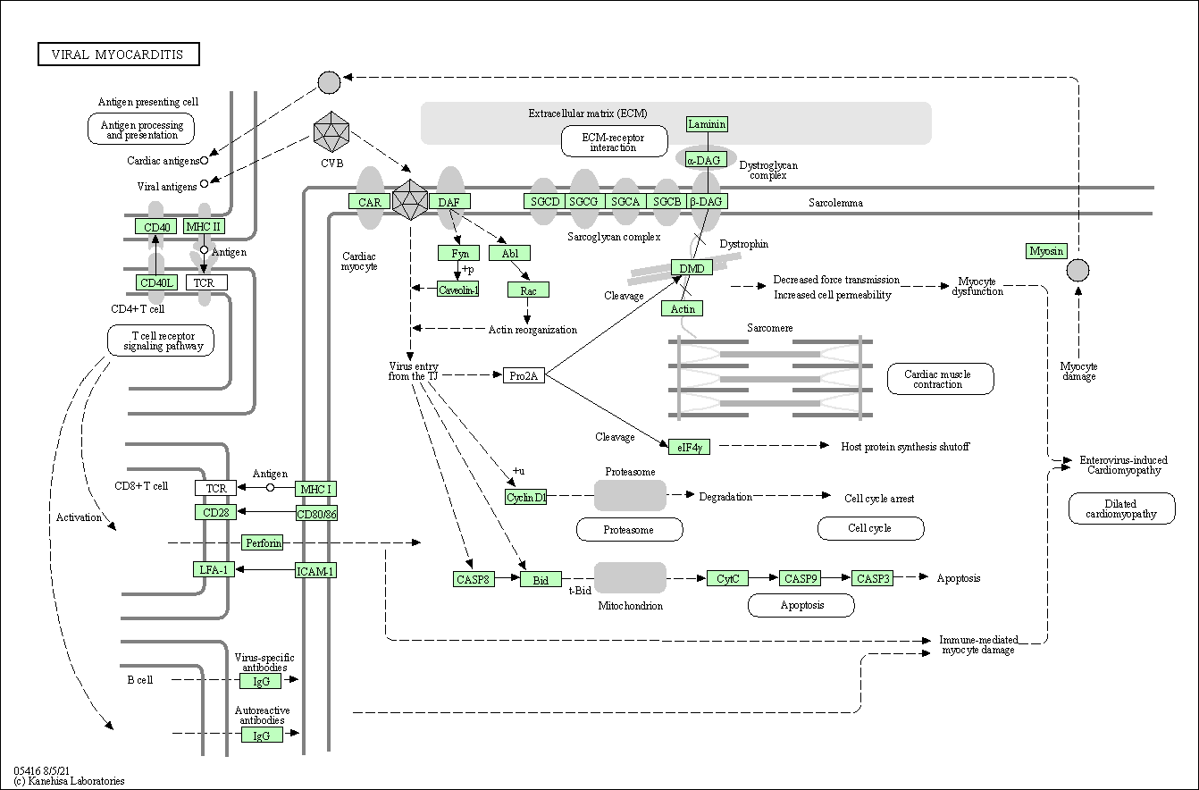
|
|||||||
| Pathogenic Escherichia coli infection | hsa05130 |
Pathway Map 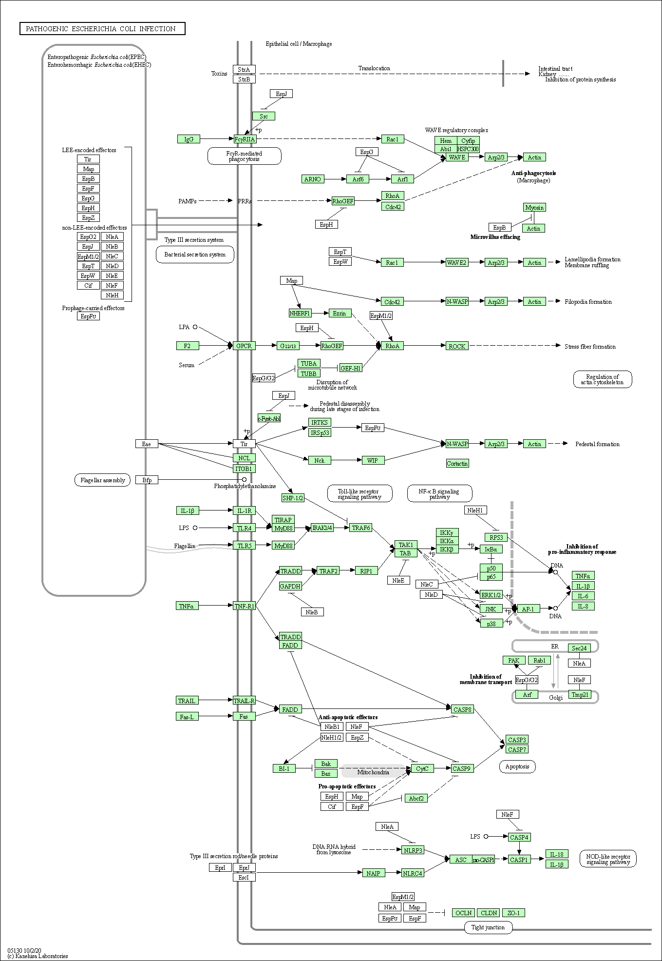
|
|||||||
| ErbB signaling pathway | hsa04012 |
Pathway Map 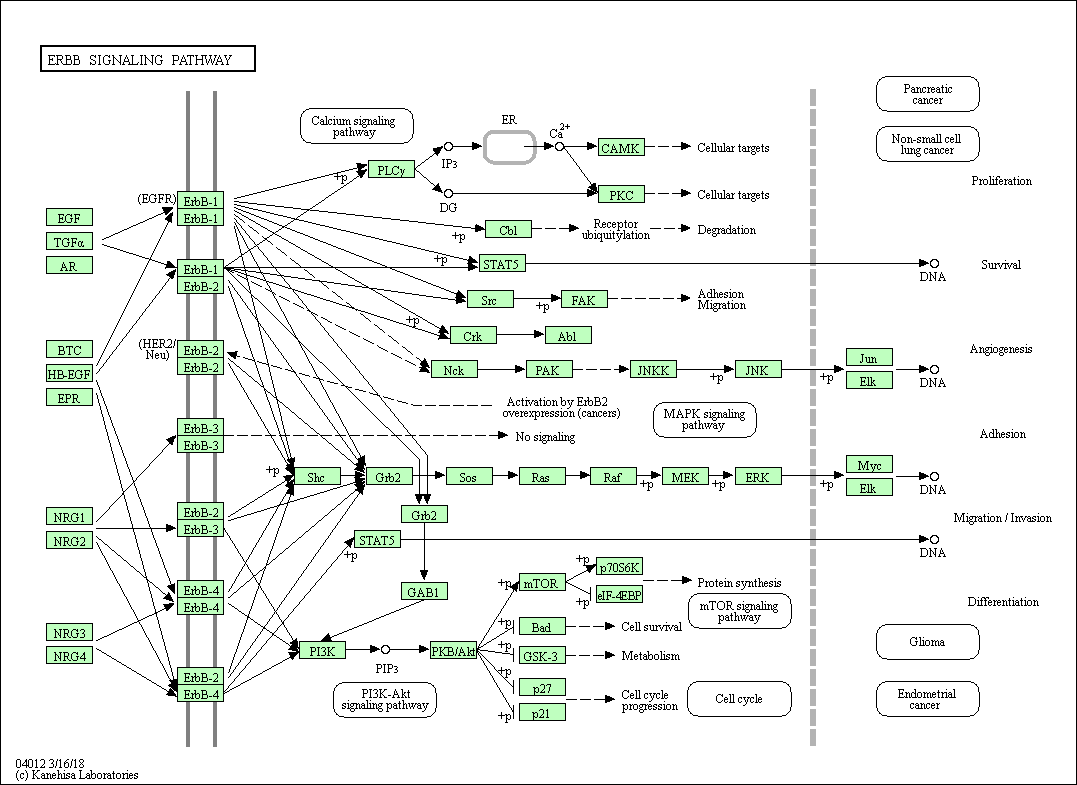
|
|||||||
| Ras signaling pathway | hsa04014 |
Pathway Map 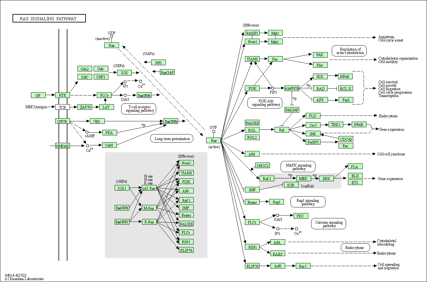
|
|||||||
| Cell cycle | hsa04110 |
Pathway Map 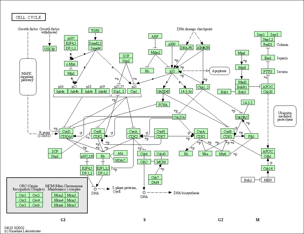
|
|||||||
| Pathways in cancer | hsa05200 |
Pathway Map 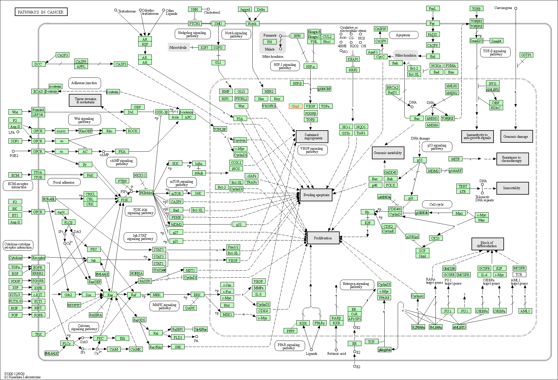
|
|||||||
| MicroRNAs in cancer | hsa05206 |
Pathway Map 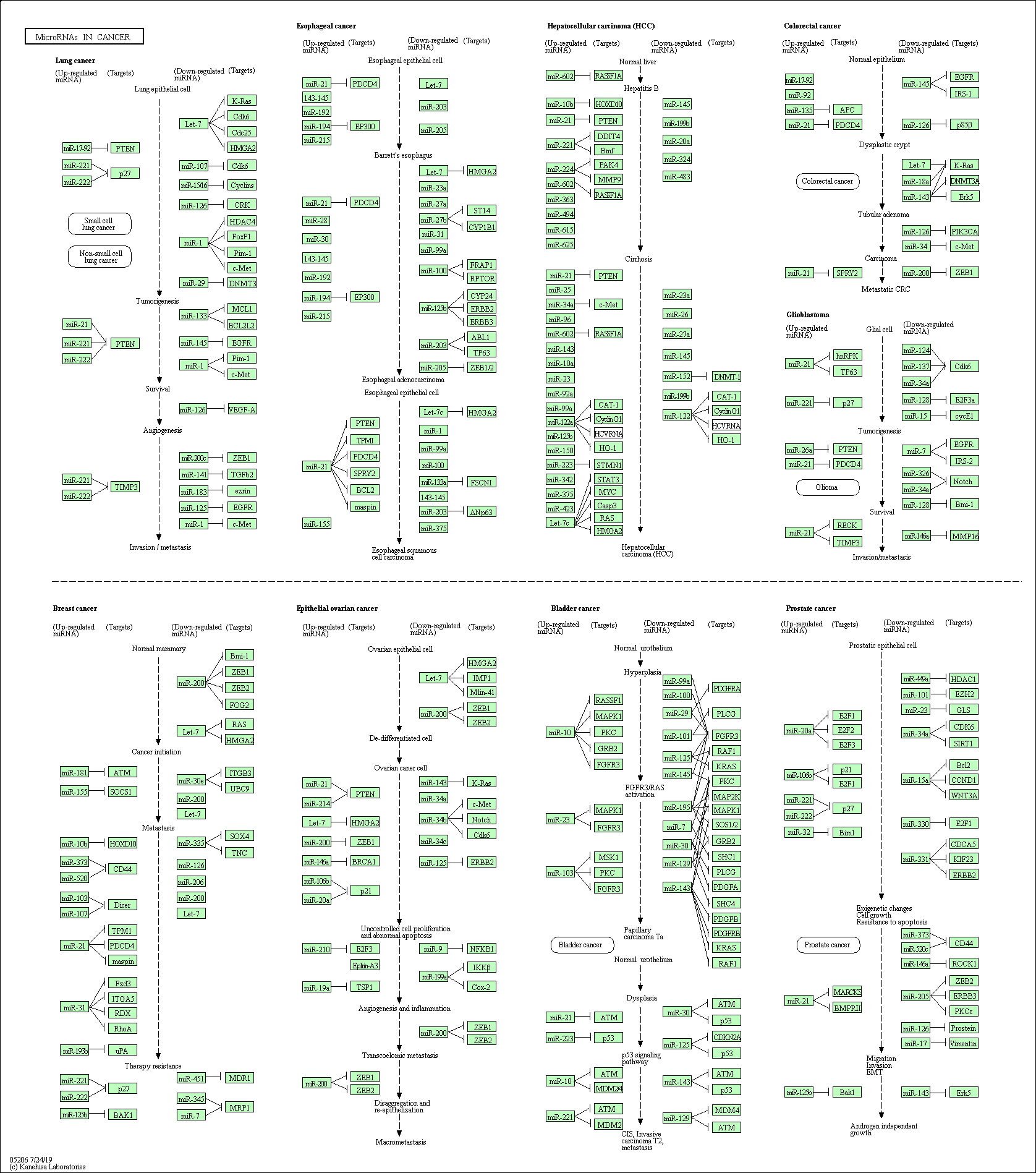
|
|||||||
| Chemical carcinogenesis - reactive oxygen species | hsa05208 |
Pathway Map 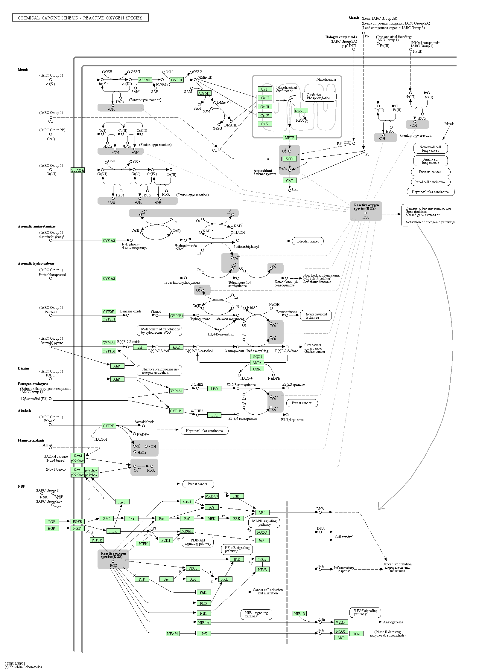
|
|||||||
| Chronic myeloid leukemia | hsa05220 |
Pathway Map 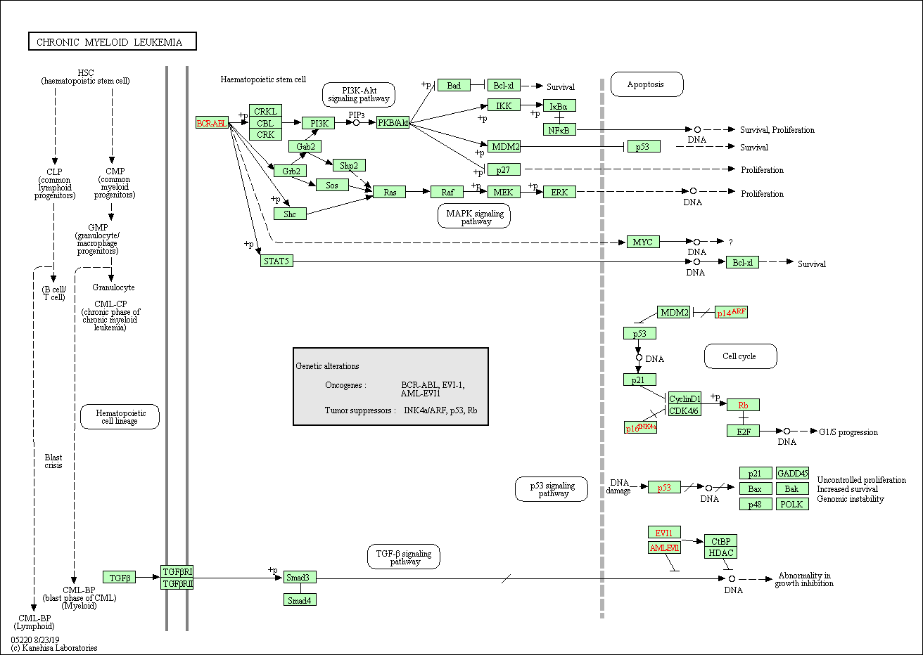
|
|||||||
| Pathogenic Escherichia coli infection | hsa05130 |
Pathway Map
|
|||||||
| ErbB signaling pathway | hsa04012 |
Pathway Map
|
|||||||
| Ras signaling pathway | hsa04014 |
Pathway Map
|
|||||||
| Cell cycle | hsa04110 |
Pathway Map
|
|||||||
| Pathways in cancer | hsa05200 |
Pathway Map
|
|||||||
| MicroRNAs in cancer | hsa05206 |
Pathway Map
|
|||||||
| Chemical carcinogenesis - reactive oxygen species | hsa05208 |
Pathway Map
|
|||||||
| 3D Structure |
|
||||||||
| Function of This Protein During Virus Infection | |||||||||
|---|---|---|---|---|---|---|---|---|---|
| Virus Name | SARS-COV-2 | Protein Function | Anti-viral | [24] | |||||
| Infected Tissue | Lung | Infection Time | 7-9 Days | ||||||
| Infected Cell | Calu-3 Cells (Human epithelial cell line) | Cellosaurus ID | CVCL_0609 | ||||||
| Method Description | To detect the role of host protein ABl1 in viral infection, ABl1 protein knockdown Calu-3 Cells were infected with SARS-COV-2 for 7 - 9 Days , and the effects on infection was detected through CRISPR-based genome-wide gene-knockout screen. | ||||||||
| Results | It is reported that knockout of ABl1 increases SARS-CoV-2 RNA levels compared with control group. | ||||||||
| Full List of Virus RNA Interacting with This Protien | |||||||||
|---|---|---|---|---|---|---|---|---|---|
| RNA Region: ORF10 (hCoV-19/Not Specified Virus Strain ) | |||||||||
| RNA Region Details |
RNA Info
 Click to show the detail information of this RNA binding region
|
[25] | |||||||
| Strains Name |
hCoV-19/Not Specified Virus Strain
|
||||||||
| RNA Binding Region |
ORF10
|
||||||||
| Virus Name |
Severe acute respiratory syndrome coronavirus 2 (SARS-CoV-2)
|
||||||||
| Infection Cells | Calu-3 cells (Human Lung Cancer Cell) (CVCL_0609 ) | ||||||||
| Cell Originated Tissue | Liver | ||||||||
| Interaction Score | P-value < 0.05 | ||||||||
| Method Description | RNA pull-down assays; liquid chromatography with tandem mass spectrometry (LC-MS/MS); Wilcoxon test; MS2 affinity purification coupled with liquid chromatography-mass spectrometry (MAMS) | ||||||||
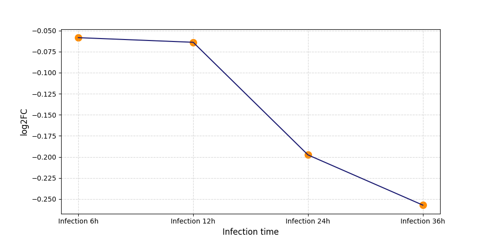
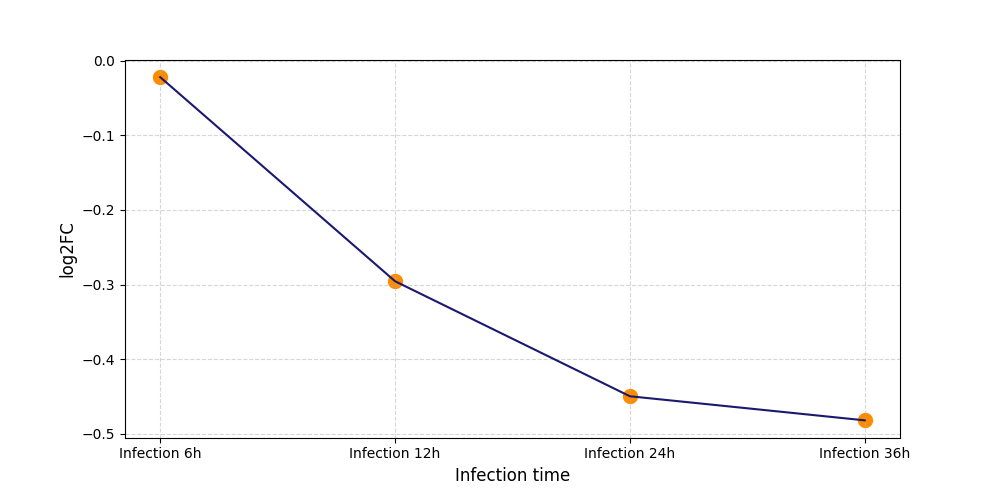
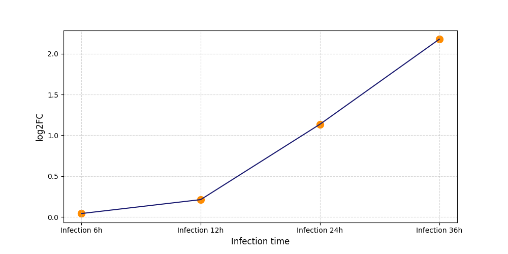
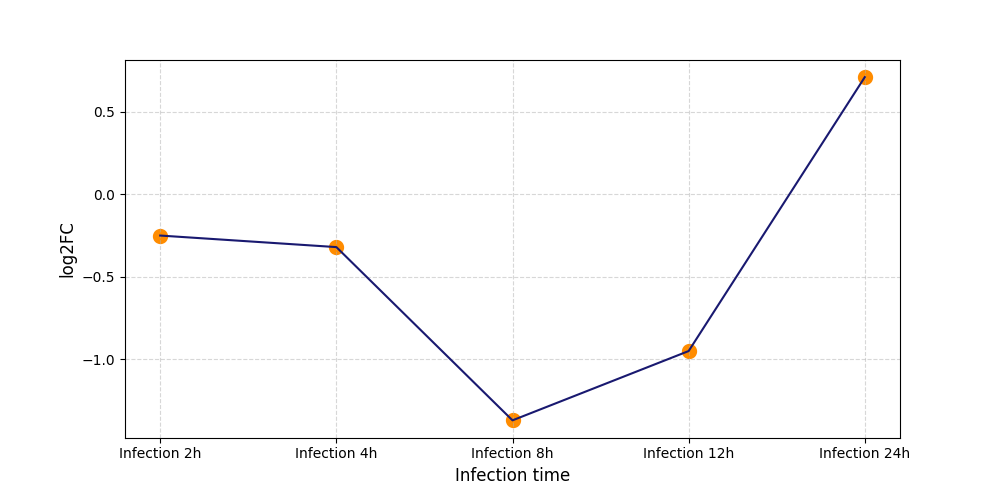
| Differential Gene Expression During SARS-COV-2 Infection | |||||||||||||||||||||||||||||||||||||||||||||||||||||||||||||||||||||||||||||||||||||||||||||||||||
|---|---|---|---|---|---|---|---|---|---|---|---|---|---|---|---|---|---|---|---|---|---|---|---|---|---|---|---|---|---|---|---|---|---|---|---|---|---|---|---|---|---|---|---|---|---|---|---|---|---|---|---|---|---|---|---|---|---|---|---|---|---|---|---|---|---|---|---|---|---|---|---|---|---|---|---|---|---|---|---|---|---|---|---|---|---|---|---|---|---|---|---|---|---|---|---|---|---|---|---|
|
|||||||||||||||||||||||||||||||||||||||||||||||||||||||||||||||||||||||||||||||||||||||||||||||||||
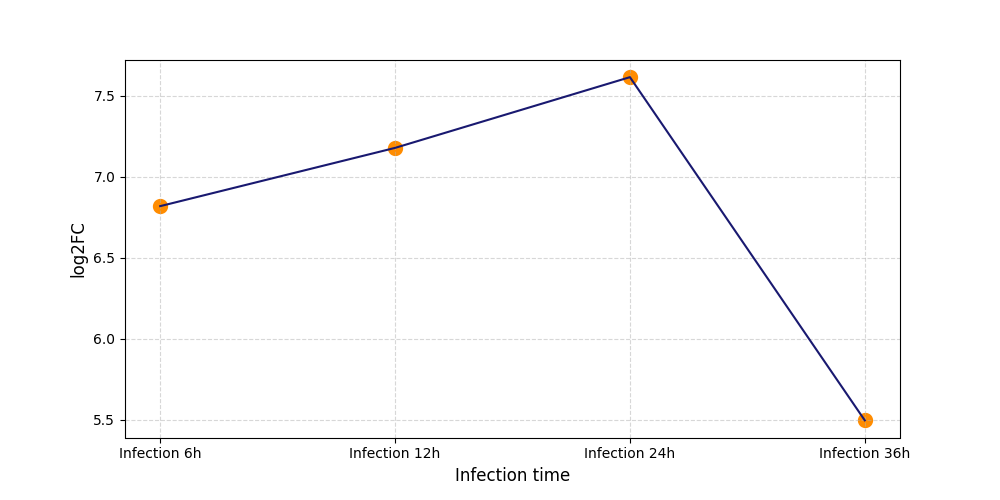
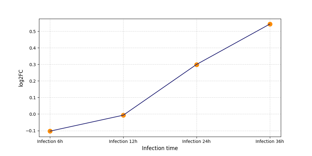
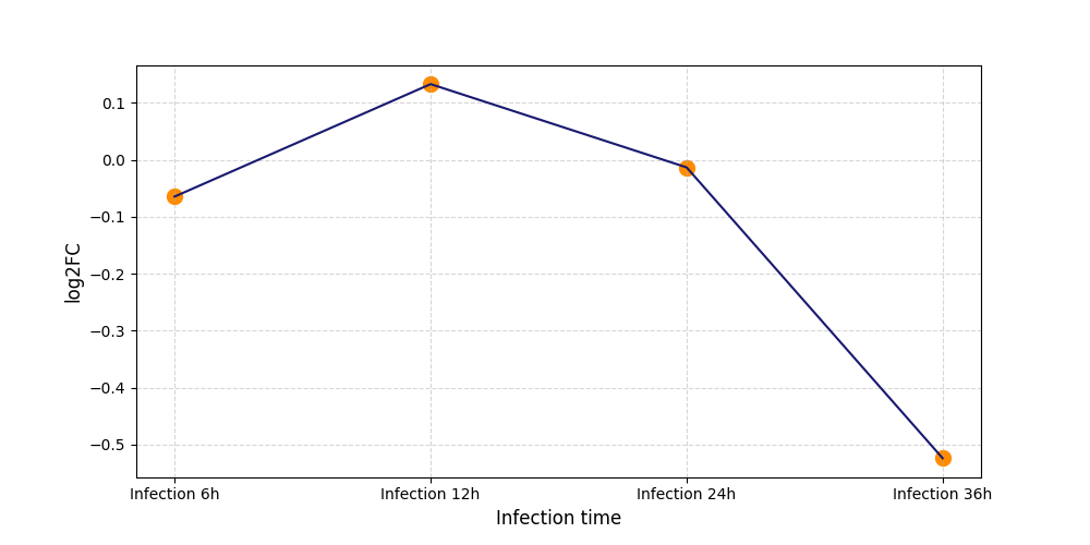
| Protein Phosphorylation after Virus Infection | |||||||||||||||||||||||||||||||||||||||||||||||||||||||||||||||||||||||||||||||||||||||||||||||||||
|---|---|---|---|---|---|---|---|---|---|---|---|---|---|---|---|---|---|---|---|---|---|---|---|---|---|---|---|---|---|---|---|---|---|---|---|---|---|---|---|---|---|---|---|---|---|---|---|---|---|---|---|---|---|---|---|---|---|---|---|---|---|---|---|---|---|---|---|---|---|---|---|---|---|---|---|---|---|---|---|---|---|---|---|---|---|---|---|---|---|---|---|---|---|---|---|---|---|---|---|
|
S569
[26] |
 |
||||||||||||||||||||||||||||||||||||||||||||||||||||||||||||||||||||||||||||||||||||||||||||||||||
|
S683
[26] |
 |
||||||||||||||||||||||||||||||||||||||||||||||||||||||||||||||||||||||||||||||||||||||||||||||||||
|
S716
[26] |
 |
||||||||||||||||||||||||||||||||||||||||||||||||||||||||||||||||||||||||||||||||||||||||||||||||||
|
S718
[27] |
 |
||||||||||||||||||||||||||||||||||||||||||||||||||||||||||||||||||||||||||||||||||||||||||||||||||
|
S718
[26] |
 |
||||||||||||||||||||||||||||||||||||||||||||||||||||||||||||||||||||||||||||||||||||||||||||||||||
|
S919
[26] |
 |
||||||||||||||||||||||||||||||||||||||||||||||||||||||||||||||||||||||||||||||||||||||||||||||||||
|
T781
[26] |
 |
||||||||||||||||||||||||||||||||||||||||||||||||||||||||||||||||||||||||||||||||||||||||||||||||||
| Potential Drug(s) that Targets This Protein | |||||||||||||||||||||||||||||||||||||||||||||||||||||||||||||||||||||||||||||||||||||||||||||||||||
|---|---|---|---|---|---|---|---|---|---|---|---|---|---|---|---|---|---|---|---|---|---|---|---|---|---|---|---|---|---|---|---|---|---|---|---|---|---|---|---|---|---|---|---|---|---|---|---|---|---|---|---|---|---|---|---|---|---|---|---|---|---|---|---|---|---|---|---|---|---|---|---|---|---|---|---|---|---|---|---|---|---|---|---|---|---|---|---|---|---|---|---|---|---|---|---|---|---|---|---|
| Drug Name | DrunkBank ID | Pubchem ID | TTD ID | REF | |||||||||||||||||||||||||||||||||||||||||||||||||||||||||||||||||||||||||||||||||||||||||||||||
| Bafetinib | DB11851 | 11387605 | D0G0VV | [27] | |||||||||||||||||||||||||||||||||||||||||||||||||||||||||||||||||||||||||||||||||||||||||||||||
| Dasatinib | DB01254 | 3062316 | D0E6XR | [27] | |||||||||||||||||||||||||||||||||||||||||||||||||||||||||||||||||||||||||||||||||||||||||||||||
| Fostamatinib | DB12010 | 11671467 | D0V8HJ | [28] | |||||||||||||||||||||||||||||||||||||||||||||||||||||||||||||||||||||||||||||||||||||||||||||||
| Imatinib | DB00619 | 5291 | D0AZ3C | [29] | |||||||||||||||||||||||||||||||||||||||||||||||||||||||||||||||||||||||||||||||||||||||||||||||
| Nilotinib | DB04868 | 644241 | . | [30] | |||||||||||||||||||||||||||||||||||||||||||||||||||||||||||||||||||||||||||||||||||||||||||||||
| Protein Sequence Information |
MLEICLKLVGCKSKKGLSSSSSCYLEEALQRPVASDFEPQGLSEAARWNSKENLLAGPSENDPNLFVALYDFVASGDNTLSITKGEKLRVLGYNHNGEWCEAQTKNGQGWVPSNYITPVNSLEKHSWYHGPVSRNAAEYLLSSGINGSFLVRESESSPGQRSISLRYEGRVYHYRINTASDGKLYVSSESRFNTLAELVHHHSTVADGLITTLHYPAPKRNKPTVYGVSPNYDKWEMERTDITMKHKLGGGQYGEVYEGVWKKYSLTVAVKTLKEDTMEVEEFLKEAAVMKEIKHPNLVQLLGVCTREPPFYIITEFMTYGNLLDYLRECNRQEVNAVVLLYMATQISSAMEYLEKKNFIHRDLAARNCLVGENHLVKVADFGLSRLMTGDTYTAHAGAKFPIKWTAPESLAYNKFSIKSDVWAFGVLLWEIATYGMSPYPGIDLSQVYELLEKDYRMERPEGCPEKVYELMRACWQWNPSDRPSFAEIHQAFETMFQESSISDEVEKELGKQGVRGAVSTLLQAPELPTKTRTSRRAAEHRDTTDVPEMPHSKGQGESDPLDHEPAVSPLLPRKERGPPEGGLNEDERLLPKDKKTNLFSALIKKKKKTAPTPPKRSSSFREMDGQPERRGAGEEEGRDISNGALAFTPLDTADPAKSPKPSNGAGVPNGALRESGGSGFRSPHLWKKSSTLTSSRLATGEEEGGGSSSKRFLRSCSASCVPHGAKDTEWRSVTLPRDLQSTGRQFDSSTFGGHKSEKPALPRKRAGENRSDQVTRGTVTPPPRLVKKNEEAADEVFKDIMESSPGSSPPNLTPKPLRRQVTVAPASGLPHKEEAGKGSALGTPAAAEPVTPTSKAGSGAPGGTSKGPAEESRVRRHKHSSESPGRDKGKLSRLKPAPPPPPAASAGKAGGKPSQSPSQEAAGEAVLGAKTKATSLVDAVNSDAAKPSQPGEGLKKPVLPATPKPQSAKPSGTPISPAPVPSTLPSASSALAGDQPSSTAFIPLISTRVSLRKTRQPPERIASGAITKGVVLDSTEALCLAISRNSEQMASHSAVLEAGKNLYTFCVSYVDSIQQMRNKFAFREAINKLENNLRELQICPATAGSGPAATQDFSKLLSSVKEISDIVQR
Click to Show/Hide
|
|---|